The Growing Importance of Musculoskeletal Ultrasound Imaging in Fishers
Musculoskeletal Ultrasound Imaging Fishers has become an essential diagnostic tool for evaluating injuries and conditions affecting muscles, joints, tendons, and ligaments. As healthcare technology continues to advance, ultrasound imaging has emerged as one of the most effective ways to observe soft tissues without the need for invasive procedures. This method uses high frequency sound waves to create real time images of structures inside the body, allowing medical professionals to analyze movement and tissue health with remarkable accuracy.
In a community like Fishers, where many residents maintain active lifestyles, musculoskeletal injuries are relatively common. Whether the issue comes from sports activities, exercise routines, or repetitive motion at work, accurate diagnosis plays a crucial role in recovery. Musculoskeletal Ultrasound Imaging Fishers allows physicians to detect injuries early and identify the precise location of damage.
The ability to visualize tissues in motion makes ultrasound imaging especially valuable. Physicians can observe tendons and muscles as they move, which helps detect abnormalities that might not appear in static imaging methods. This dynamic capability makes ultrasound one of the most versatile diagnostic tools available today.
Another advantage of this technology is accessibility. Many clinics and medical imaging centers in Fishers offer ultrasound services because the equipment is portable and efficient. Patients often receive quick appointments and fast results, reducing the stress associated with waiting for a diagnosis.
Ultrasound imaging also enhances communication between healthcare providers and patients. Since images appear instantly on a monitor, physicians can explain findings in real time. This helps patients better understand their condition and the steps needed for recovery.
Because of these benefits, Musculoskeletal Ultrasound Imaging Fishers continues to play a growing role in modern healthcare. It supports early diagnosis, guides treatment decisions, and helps patients return to their normal activities more quickly.
How Ultrasound Technology Helps Diagnose Musculoskeletal Conditions
Ultrasound technology works by sending high frequency sound waves into the body using a device known as a transducer. These sound waves travel through tissues and reflect back when they encounter different structures. The ultrasound machine then converts these reflected waves into detailed images that appear on a screen.
Different types of tissue reflect sound waves in unique ways, which allows physicians to distinguish between muscles, tendons, ligaments, and fluid filled spaces. This capability makes Musculoskeletal Ultrasound Imaging Fishers particularly useful for evaluating soft tissue injuries.
One of the most valuable aspects of ultrasound imaging is real time visualization. Unlike some imaging techniques that capture a single snapshot, ultrasound allows continuous observation of tissues as they move. Physicians can watch how joints function and how tendons glide across surrounding structures.
This real time evaluation is extremely helpful when diagnosing conditions related to movement. For example, a tendon may appear normal when the joint is at rest but show abnormalities when the patient moves the affected area. Ultrasound imaging makes it possible to capture these changes during the scan.
Another strength of ultrasound is the ability to examine very small structures with high precision. Modern machines produce high resolution images that reveal subtle changes in tissue texture and shape. These details help physicians detect injuries early and recommend appropriate treatment.
The non invasive nature of the procedure also makes it suitable for repeated monitoring. Physicians can perform follow up scans to evaluate healing progress or monitor chronic conditions over time.
Through its combination of accuracy, speed, and safety, Musculoskeletal Ultrasound Imaging Fishers has become a trusted diagnostic solution for many healthcare providers.
Musculoskeletal Injuries and Conditions Commonly Detected With Ultrasound
Musculoskeletal ultrasound is widely used to identify a variety of injuries and medical conditions affecting soft tissues. Many patients seek imaging after experiencing pain, swelling, stiffness, or reduced mobility in a joint or muscle.
Tendon injuries are one of the most common findings during ultrasound examinations. Tendinitis occurs when tendons become inflamed due to overuse or strain. Ultrasound can reveal thickened tendons, irregular fiber patterns, or small tears that indicate injury.
Muscle injuries also appear clearly on ultrasound images. Athletes and physically active individuals often develop muscle strains or partial tears after intense activity. Ultrasound helps determine the severity of the damage and assists physicians in designing rehabilitation plans.
Joint related conditions can also be evaluated using Musculoskeletal Ultrasound Imaging Fishers. Inflammation within the joint may indicate arthritis or other inflammatory conditions. Ultrasound scans reveal fluid accumulation, synovial swelling, and structural changes within the joint.
Ligament injuries are another frequent reason for musculoskeletal imaging. Sprains and ligament tears can cause pain and instability around joints such as the ankle or knee. Ultrasound helps physicians determine whether the ligament is partially damaged or completely torn.
Nerve compression conditions can also be identified during ultrasound examinations. When nerves become compressed by surrounding tissues, patients may experience numbness, tingling, or weakness. Ultrasound allows physicians to visualize the affected nerve and identify the source of compression.
By detecting these conditions early, Musculoskeletal Ultrasound Imaging Fishers helps patients begin treatment sooner and avoid further complications.
Benefits of Musculoskeletal Ultrasound Imaging for Patients
Musculoskeletal ultrasound imaging provides several advantages that make it appealing for both patients and healthcare professionals. One of the most important benefits is the comfort of the procedure. Patients simply lie down or sit comfortably while the physician moves a small handheld device across the skin.
The scan does not involve radiation exposure, which is a major advantage compared to certain imaging methods. Because ultrasound uses sound waves instead of radiation, it can be performed multiple times without health risks.
Another significant benefit is the speed of the examination. Many musculoskeletal ultrasound scans take less than thirty minutes to complete. This efficiency allows patients to receive diagnostic results quickly and move forward with treatment.
Cost effectiveness is also an important consideration. Ultrasound imaging is generally more affordable than some other advanced imaging procedures. This accessibility makes Musculoskeletal Ultrasound Imaging Fishers a practical choice for individuals seeking diagnostic evaluation.
Real time imaging adds another level of diagnostic accuracy. Physicians can observe how muscles and tendons move during the scan, helping them detect issues that might not appear in static images.
Key Advantages Patients Experience
Musculoskeletal ultrasound imaging offers several practical benefits that improve the diagnostic experience for patients.
• Non invasive imaging with minimal discomfort
• No radiation exposure during the procedure
• Real time visualization of joints and soft tissues
• Quick appointment times and shorter scan durations
• Accurate evaluation of muscles, tendons, ligaments, and joints
• Lower cost compared to many advanced imaging methods
• Useful for guiding injections and other treatments
These advantages highlight why Musculoskeletal Ultrasound Imaging Fishers continues to gain popularity among patients and healthcare providers.
The Role of Ultrasound Imaging in Sports Medicine
Sports medicine specialists frequently rely on ultrasound imaging to diagnose injuries in athletes. Many sports injuries involve soft tissues, making ultrasound an ideal diagnostic tool. Musculoskeletal Ultrasound Imaging Fishers allows physicians to evaluate damage quickly and accurately.
Athletes often experience overuse injuries due to repetitive movements during training. Tendons and muscles may develop small tears or inflammation that gradually worsen over time. Ultrasound imaging can detect these problems early, allowing athletes to adjust their training routines before the injury becomes severe.
Dynamic imaging is especially valuable in sports medicine. Physicians can observe how tendons and joints behave while the athlete performs simple movements. This helps identify mechanical problems that contribute to pain or instability.
Ultrasound imaging also assists with monitoring recovery. After an injury occurs, physicians can perform follow up scans to track the healing process. This helps determine when an athlete can safely return to training or competition.
Sports medicine clinics in Fishers often keep portable ultrasound equipment on site. This allows physicians to evaluate injuries immediately without sending athletes to a separate imaging facility.
Because of its efficiency and accuracy, musculoskeletal ultrasound has become a vital tool for protecting the health and performance of athletes.
What Happens During a Musculoskeletal Ultrasound Scan
Patients scheduled for Musculoskeletal Ultrasound Imaging Fishers often appreciate how simple the process is. Preparation typically requires little effort. Most patients only need to wear clothing that allows access to the area being examined.
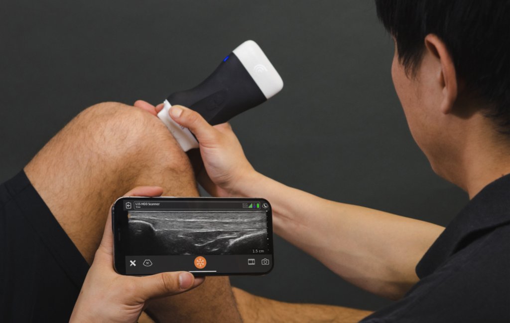
At the beginning of the scan, the technician or physician applies a small amount of gel to the skin. This gel allows the ultrasound transducer to glide smoothly and improves sound wave transmission.
The transducer is then moved across the surface of the skin while the machine captures images of the underlying tissues. The physician may ask the patient to move the joint or muscle slightly to observe its motion in real time.
The images appear immediately on a monitor, allowing the healthcare provider to analyze them during the examination. This instant feedback helps identify abnormalities quickly.
Most musculoskeletal ultrasound scans take between fifteen and thirty minutes depending on the complexity of the condition being evaluated.
After the scan, the physician discusses the findings with the patient and recommends the next steps. These steps may include physical therapy, medication, or further imaging if necessary.
Individuals Who Benefit Most From Musculoskeletal Ultrasound Imaging
Many different groups of people benefit from musculoskeletal ultrasound imaging. Active adults who participate in sports or regular exercise often rely on this technology to diagnose injuries quickly.
Athletes in particular benefit from Musculoskeletal Ultrasound Imaging Fishers because the scan provides immediate information about muscle and tendon injuries. This allows trainers and physicians to develop rehabilitation plans without delay.
Individuals experiencing chronic joint pain may also benefit from ultrasound imaging. Conditions such as arthritis can cause inflammation within the joint that is easily visible on ultrasound scans.
Older adults often experience degenerative changes in the musculoskeletal system. Ultrasound imaging helps monitor these conditions and guides treatment decisions aimed at improving mobility and comfort.
Workers who perform repetitive tasks may develop strain injuries in areas like the wrist, elbow, or shoulder. Ultrasound scans can identify tissue damage caused by repetitive movement and help determine appropriate therapy.
Because it is safe and versatile, musculoskeletal ultrasound can be used for patients of various ages and activity levels.
Ultrasound Guided Treatments and Procedures
Musculoskeletal ultrasound is not limited to diagnosis alone. Physicians frequently use this imaging technology to guide therapeutic procedures with greater precision. During these procedures, ultrasound allows the physician to watch the needle as it moves toward the targeted area.
One of the most common ultrasound guided treatments involves corticosteroid injections. These injections reduce inflammation and relieve pain in joints or tendons. Ultrasound guidance ensures the medication reaches the exact location where it is needed.
Platelet rich plasma therapy is another treatment often guided by ultrasound imaging. This therapy uses the patient’s own blood components to stimulate healing in damaged tissues.
Ultrasound guidance also reduces the likelihood of complications during procedures. Physicians can clearly see surrounding structures such as nerves and blood vessels, helping them avoid accidental injury.
Because of the precision offered by ultrasound guidance, patients often experience improved treatment results and faster recovery.
Advancements Shaping the Future of Musculoskeletal Ultrasound
The field of ultrasound imaging continues to evolve rapidly as new technologies improve image quality and diagnostic capabilities. Modern ultrasound systems produce highly detailed images that allow physicians to detect even minor abnormalities.
Portable ultrasound machines have expanded the availability of Musculoskeletal Ultrasound Imaging Fishers. These devices enable physicians to perform scans in clinics, sports facilities, and emergency departments.
Artificial intelligence is also beginning to influence diagnostic imaging. Some ultrasound systems now include software that assists physicians in identifying patterns and abnormalities within the images.
Improved real time image processing allows for smoother visualization of moving structures. This advancement enhances the ability to study joint movement and tendon function.
As these innovations continue to develop, musculoskeletal ultrasound will likely become even more accurate and widely available.
Takeaway
Musculoskeletal Ultrasound Imaging Fishers offers a reliable and efficient way to evaluate injuries affecting muscles, joints, tendons, and ligaments. The technology provides real time images that help physicians detect problems quickly and develop targeted treatment plans. Patients benefit from a safe, non invasive procedure that delivers accurate diagnostic information without radiation exposure. Whether addressing sports injuries, chronic pain, or mobility issues, ultrasound imaging plays a vital role in modern musculoskeletal care. As technology continues to advance, this diagnostic method will remain an important resource for healthcare providers and patients seeking effective solutions for soft tissue conditions.
Frequently Asked Questions
What does Musculoskeletal Ultrasound Imaging Fishers diagnose
It helps diagnose injuries affecting muscles, tendons, ligaments, joints, and nerves, including inflammation, tears, and fluid buildup.
Is the ultrasound scan painful
The scan is generally painless. Patients may only feel mild pressure from the transducer as it moves across the skin.
How long does a musculoskeletal ultrasound appointment take
Most appointments last between fifteen and thirty minutes depending on the area being examined.
Can ultrasound detect ligament injuries
Yes. Musculoskeletal ultrasound can identify sprains, partial tears, and other ligament abnormalities.
Is ultrasound imaging safe for repeated use
Yes. Because it does not use radiation, ultrasound imaging can be safely repeated whenever necessary.
Do athletes commonly use musculoskeletal ultrasound
Yes. Sports medicine physicians frequently use ultrasound to diagnose and monitor sports related injuries.
Can ultrasound guide injections and treatments
Yes. Physicians often use ultrasound imaging to guide injections and ensure accurate placement of medications.